How a 3D/4D Ultrasound Works: The Science Behind the Magic
Ever wonder how 4d ultrasound works? Discover the fascinating science of sound waves, echoes, and how our studio turns them into beautiful baby portraits.


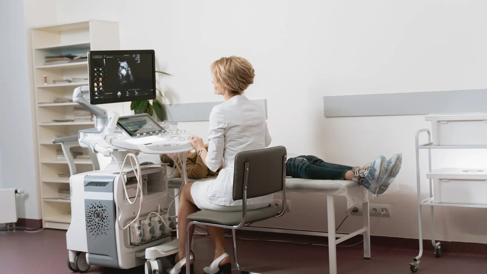
When you lie back in our comfortable studio chairs, watch the lights dim, and suddenly see your baby yawning on a 70-inch screen, it truly feels like magic. But behind that emotional, tear-jerking moment is decades of incredible physics and computer science. If you have ever asked yourself how 4d ultrasound works, you are in for a treat.
At Baby Studio 4D, we think the science is just as beautiful as the images. Understanding the technology not only satisfies your curiosity but also helps explain why it is so safe for your growing baby. Let’s pull back the curtain and explore how we turn invisible sound into unforgettable memories.
(Note: Elective ultrasounds are for bonding and keepsake purposes and do not replace diagnostic medical evaluations from your OB-GYN.)
The Basics: Echolocation (Like a Dolphin!)
The core technology behind every single ultrasound—whether it’s a grainy 2D scan at your doctor’s office or a hyper-realistic 8K portrait at our studio—is the exact same: sound waves.
Inside the ultrasound wand (the transducer), there are tiny piezoelectric crystals. When an electrical current hits these crystals, they vibrate rapidly, creating high-frequency sound waves. These sound waves are pitched so high that human ears cannot hear them.
When we place the wand on your belly, these sound waves travel through the amniotic fluid until they hit something solid—like your baby’s nose or hand. When the wave hits the baby, it bounces back (echoes) to the wand. To understand how the quality of these waves affects your image, you can read our article on the evolution of ultrasound technology.
Translating Sound into Sight
How does an echo become a picture? The ultrasound machine is basically a supercomputer with a stopwatch.
It calculates exactly how long it took for the sound wave to travel down, hit the baby, and bounce back. If it bounces back quickly, the machine knows the object is close to the surface. If it takes longer, the object is deeper. By sending and receiving millions of these pulses per second, the computer maps out the exact shape and location of everything inside the womb.
If you are curious about how this fundamental science relates to safety, we cover it thoroughly in our post exploring whether elective ultrasounds are safe.
From 2D to 3D: Slicing the Image
Standard 2D ultrasound sends waves in a single, straight line, producing a flat cross-section. So how do we get a 3D face?
Instead of looking straight down, the crystals inside a 3D transducer automatically sweep back and forth mechanically within the wand housing. This sends sound waves at hundreds of slightly different angles. The computer then takes all these 2D “slices” and stitches them together, creating a three-dimensional volume. It’s like taking a loaf of bread, slicing it, and then putting it back together to see the whole crust!
For a more detailed breakdown of how these different dimensions compare visually for parents, read our 2D vs 3D vs 4D vs HD comparison guide.
Adding the 4th Dimension: Time
This is where the magic really happens. 4D ultrasound isn’t a new type of space; it is simply 3D images updated in real-time.
Our powerful studio computers process those 3D volumes so incredibly fast (several times per second) that it creates a live video feed. This is what allows you to watch your baby kick, smile, or suck their thumb live on the screen. It requires immense processing power, which is why specialized studios like ours invest heavily in top-tier machines as detailed in our main guide to modern ultrasounds.
Conclusion
The next time you see an ultrasound image of your baby, you’ll know that you are actually looking at millions of tiny, silent echoes perfectly translated into a photograph by a supercomputer. It is a brilliant blend of biology, physics, and art.
Ready to see the science in action? Book your 3D/4D ultrasound session at Baby Studio 4D and let us introduce you to your baby!
Frequently Asked Questions
Frequently Asked
Does the ultrasound machine use radiation?
Why does the technician use that cold gel?
How does the machine know what color to make the baby?
Can the baby hear the ultrasound waves?
Every milestone
deserves to be
seen clearly.
Bronze
- 2D Ultrasound Session
- Black & White Printed Pictures
- Hear Baby's Heartbeat
- Baby Growth Measurements
Silver
- 2D Ultrasound Session
- Boy or Girl Reveal
- Gender Confirmation
- 3D Image (if baby cooperates)
- Digital Pictures (Momitalk)
Gold
- 3D / HD Ultrasound Session
- Gender Confirmation
- Digital Pictures (Momitalk)
- 5 Color Pictures Printed
- Baby Growth Measurements
Platinum
- 3D / 4D / 5D / HD Session
- Gender Confirmation
- Digital Video & Pictures
- 8 Color Pictures Printed
- Baby Growth Measurements
Mommy Bundle
- Gender Confirmation
- Hear Heartbeat
- 6 Color + Digital Pictures
- Unlimited Digital Video
- 8K Hyperrealistic Image
- Small Bear with Baby's Heartbeat
- Full Baby Growth Report
Gift Card
Choose a package for your loved ones, or for that special someone you want to treat.
Bilingual sonographer based in Raleigh, NC. Six years of experience guiding families through their first 3D, 4D, and 8K HD ultrasound sessions — written here as field notes from the studio.
More from the journal
Ultrasound Technology Complete Guide: From Sound Waves to 8K HD
The definitive guide to ultrasound technology for expecting parents. Understand how sound becomes images, from…

Ultrasound Frequency Explained: Why Studio Quality Matters
Not all 4D ultrasounds are equal. Learn how high quality ultrasound studio equipment and proper frequencies cr…

Medical Ultrasound vs Elective Ultrasound: Key Differences Explained
Confused about medical vs elective ultrasound? Discover the key differences in purpose, technology, and experi…
Ready to meet your baby?
Over 1,000 families have already shared their first moments with us. Yours could be next.
Book your session →